
Ultrasound: Gall Bladder Edition
Learn about chronic pain management with our CPD course!
For those of you who might not know, I had a 9 month stint in radiology. I thought it might be the career for me - sessional speciality, diagnosis focussed and a great lifestyle. I quickly realised that it probably was not the career for me as I really missed patients but I certainly learnt lots from there.
What was most intriguing is how far medical imaging has come. Most of us are acquainted with x-ray interpretation but that’s as far as our image interpretation goes. Certainly after X-rays, ultrasound imaging is the most common type of imaging. I’ve probably requested hundreds of ultrasound for abdominal pain and at times, it’s helpful to know exactly what an ultrasound can uncover so as to appropriately request imaging. I thought I’d use this weeks newsletter to showcase some ultrasound images for conditions affecting the liver and gallbladder.
Principles of ultrasound
Ultrasound works by sending high-frequency sound waves into the body using a transducer. These waves bounce off tissues and organs, creating echoes that are captured and converted into real-time images by a computer. Sonographers can alter the angles and planes (transverse vs longitudinal) to visualise various organs.
Abdominal pain is a common reason for why we perform ultrasound. Visualising the gallbladder is part of this and a monographer will measures:
- Gallbladder size and contents
- Wall thickness (to assess inflammation —> cholecystitis)
- Common Bile Duct (diameter < 7mm is normal)
Here are a series of images that illustrate common pathology:
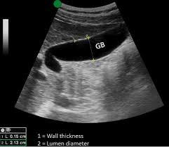
Normal gallbladder. Notice the empty lumen and thin walls.
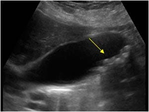
Gallstones in the gallbladder. Notice the numerous small, circular-like structure.
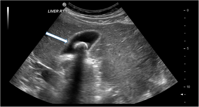
Large gallbladder calculus. A gallbladder calculus of this size will almost certainly explain a patients abdominal pain. The wall is also slightly thickened which could either suggest overgrowth (as the gallbladder tries to push the stone out) or inflammation. How does a sonographer differentiate this?
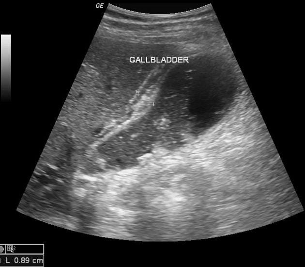
Ultrasound showing some gallstones and gallbladder wall thickening. This thickening is secondary to inflammation which is evidenced by the pericholecystic fluid (demarcated by the “+” signs).
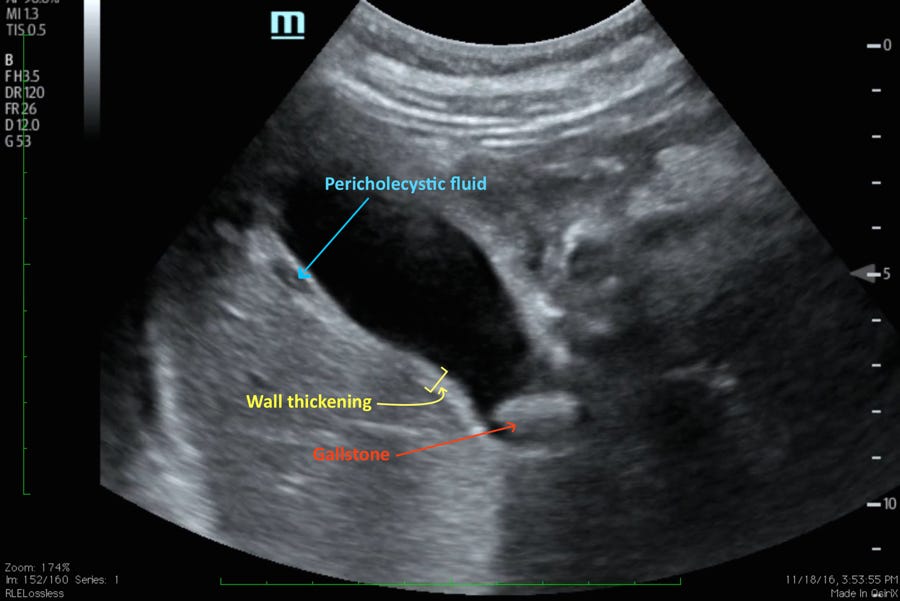
Labelled image illustrating the concepts above. This image is consistent with a diagnosis of cholecystitis.
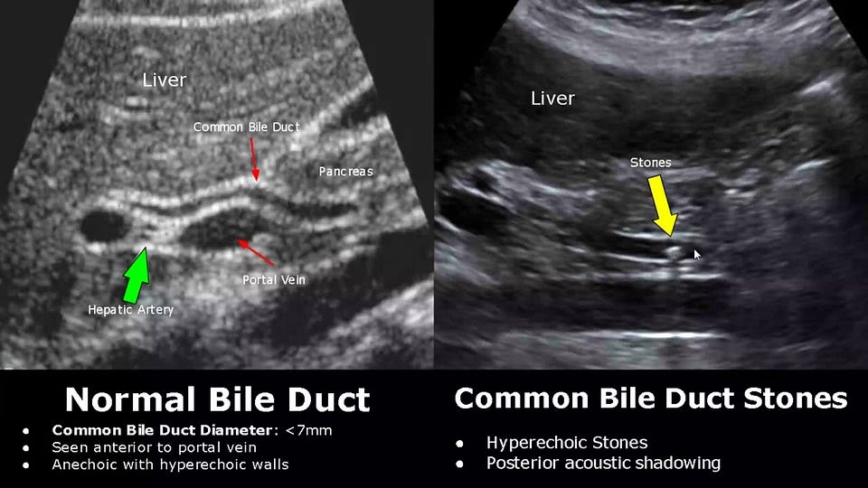
Lastly, here is an image of the common bile duct. Notice the thin structure of the CBD which is < 7mm especially when compared to the dilated CBD. In this image, the gallstone can be visualised but this is not always the case.
![Dilatation of the common bile duct [2 images] – EFSUMB](https://substackcdn.com/image/fetch/$s_!nqEp!,w_1456,c_limit,f_auto,q_auto:good,fl_progressive:steep/https%3A%2F%2Fsubstack-post-media.s3.amazonaws.com%2Fpublic%2Fimages%2Fe259da19-05e7-45b2-ae63-df235a8273e5_1200x480.png)
Ultrasound image showing a dilated CBD superior to the portal vein.
Liver pathology on ultrasound
As well as telling us about gallbladder pathology, an ultrasound scan is also a useful way to assess the liver for both benign and serious pathology. This includes:
- Fatty liver disease
- Hepatocellular carcinoma
- Liver Cirrhosis
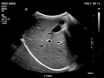
This is a normal ultrasound image of the liver. Note the slightly matt, darker echo-texture of the liver. This becomes relevant when imaging patients with fatty liver disease.
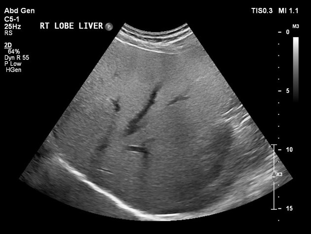
In hepatic steatosis or fatty liver, the echo-texture becomes shiny which represents the yellow/white fat deposition on the surface of the liver.
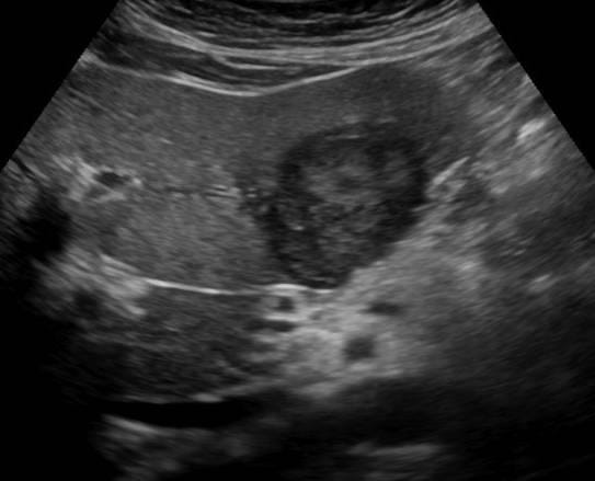
This is an ultrasound image showing an unfortunate diagnosis of hepatocellular carcinoma (HCC). It sits within the liver (as is restricted by the liver border) with some surrounding oedema (darker areas). The irregular borders also lends to a diagnosis of a HCC.
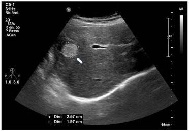
The above is contrasted with a liver haemangioma. This is a more common finding where the lesion is brighter (blood), has a regular border and there are no surrounding changes (e.g. oedema). Any diagnostic uncertainty is referred further for biopsy.
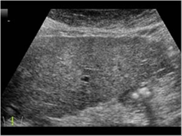
Finally, we have an ultrasound image of liver cirrhosis. Scroll up and compare this image to the normal liver ultrasound image. Notice the breakdown of the tissue and spay attention to the “bubbly border” of the liver edge.
That’s the end of our little series on liver and gallbladder ultrasound imaging. We hope that brings your ultrasound requests “to life” as we always feel that knowing what an onward referral can uncover is always helpful for a better clinical assessment and more accurate requests for investigations.
CPD Pain Management Masterclass

Finally, on the topic of pain, don’t forget to sign onto our CPD accredited Pain Management Masterclass on Sunday 2nd March 2025. It will be recorded so you will have the chance to watch again! You can read more here.
Until next time.
Website: www.paretoeducation.co.uk
Instagram: www.instagram.com/pareto_ed
Twitter: www.twitter.com/pareto_ed
Youtube: https://bit.ly/3DPm23c
Email: info@paretoeducation.co.uk
Originally published on Substack.